Tampilkan postingan dengan label Medical Training. Tampilkan semua postingan

Senin, 26 Desember 2011

Case Study: A Puzzling Facial Rash on a 17-Year-Old Boy

BACKGROUND

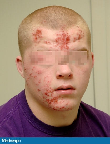
Figure 1

A 17-year-old male high school student presents to the pediatric infectious disease clinic complaining of a 10-day history of a facial rash that "won't get better." The patient had previously visited his primary care provider (PCP), who started the patient on amoxicillin-clavulanic acid 8 days ago. The rash did not improve on the antibiotic, and as a result, it was discontinued and the patient switched to trimethoprim-sulfamethoxazole. No improvement was noted with the second round of antibiotic therapy; the rash continued to spread, and the lesions increased in number. The patient was subsequently advised to follow up with the infectious disease clinic.


At the infectious disease clinic, the patient states that the rash started with several pimples over the forehead and cheek and then continued to spread and involve most of the right side of his face. The lesions are not itchy, but they are painful. The patient has no known drug allergies. His immunizations are up to date. He is very active on the wrestling team and was happily preparing for an upcoming competition. The patient denies having any weight loss, headaches, dizziness, photophobia, fever, or chills. The family history is noncontributory.
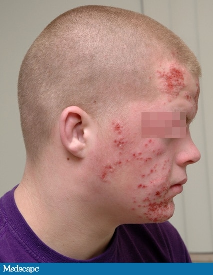

Figure 2
On physical examination, the patient is alert and orientated. His oral temperature is 97.0°F (36.1°C). The patient has normal heart sounds, his pulse has a regular rhythm of 97 bpm, and his blood pressure is 125/75 mm Hg. His lungs are clear and his respiratory rate is 12 breaths/min. The examination of the head, eyes, ears, and nose is remarkable for multiple vesicular lesions measuring about 0.5 cm in diameter (see Figures 1 and 2). There is bilateral submandibular lymph gland enlargement measuring 1.5 × 1 cm. The neck is supple. His abdomen is soft and nontender to deep palpation in the epigastric region, and no organomegaly is noted. A complete blood count (CBC) taken at the PCP's office showed a white blood cell (WBC) count of 7.4 × 10³/µL (7.4 × 10⁹/L), with a normal differential; a hemoglobin of 13.6 g/dL (136 g/L); a hematocrit of 38.3% (0.3830); and a platelet count of 298 × 103/uL (298 × 109/L).
Selasa, 06 Desember 2011

Kasus: A 44-Year-Old Man With Back Pain and Progressive Weakness

Background

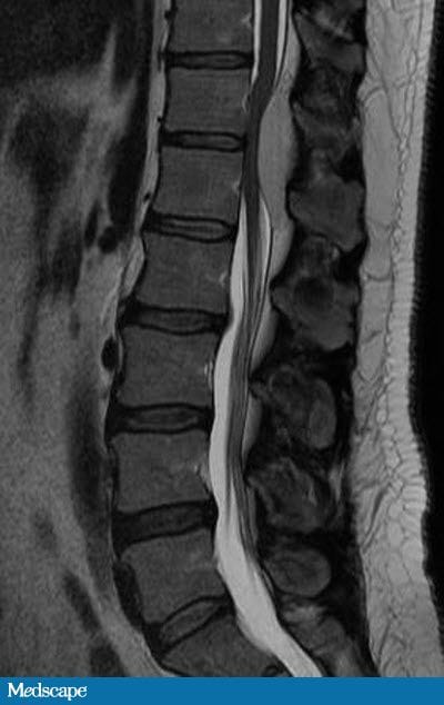
A 44-year-old man is referred for neurosurgical evaluation secondary to a 6-month history of progressive bilateral lower extremity numbness and weakness. Additionally, he reports a history of back pain over the past 4 years that he describes as an occasional sharp shooting pain down his right thigh. The only medication that he has taken is ibuprofen for pain relief. His surgical history only includes an appendectomy at 14 years of age. He denies tobacco, alcohol, or illicit drug use.
Figure 1

Kamis, 24 Maret 2011

Kasus: Sudden Onset Chest Pain in a Young Man

History

An 18-year-old man presents to the emergency department stating he felt like he “was going to die.”  While on break at work, he experienced sudden onset sharp, midsternal chest pain.  He had no dyspnea or radiation of the pain.  He had not suffered any recent chest trauma but did strike his head on a piece of equipment at work without sequelae. Three days prior to presentation he had cough productive of yellow sputum without fevers or chills.  At the time of evaluation, the pain had been present for several hours and was only relieved by narcotic medications.
His past medical history was significant for bipolar disorder, which was controlled without medications.  He worked as a stock clerk at a department store.  He was a current smoker, 1 pack per day for 2 years, and he gave a history of prior cocaine use, but denied any recent illicit drug usage.

Kamis, 10 Maret 2011

Kasus: lession on head


The patient is suffering from exophthalmos , lytic bone lesions and diabetes insipidus


WHAT IS YOUR DIAGNOSIS FOR THIS CASE???

Kasus: High creatinin and Urea after back pain medication

A 46-year-old Caucasian male (CM) with chronic back pain takes large doses of over-the-counter pain medications. He works 12-hour night shifts and has to take 15-20 pills, especially Motrin (ibuprofen) and Aleve (naproxen), during the shift and about the same amount during the day in order to "function."

He went to the ER today because of his back pain. CBC and BMP are ordered.

Past medical history (PMH)Back pain.

Medications
Motrin (ibuprofen), Aleve (naproxen), Advil (ibuprofen), Ultram (tramadol), Tylenol (acetaminophen).

Physical examination
Limited range of motion (ROM) of lower back, otherwise unremarkable.

Laboratory results
BMP showed a creatinine of 3.3 mg/dL, BUN 25 mg/dL. CBC was normal.

The patient had a normal CBC and BMP one month ago.


Author: V. Dimov, M.D.
Reviewer: S. Noor, M.D.
What is the most likely diagnosis?
What tests would your order?

Rabu, 09 Maret 2011

Wheezing dan Eosinophilia pada laki-laki umur 24 tahun

WRITE YOUR DIAGNOSIS!!!!

History

The patient is a 24-year-old Asian-Indian male who initially presented to a community hospital with a 10-day history of mild hemoptysis (2 -3 tablespoons of blood per day), followed by arthralgias, joint stiffness (knees, shoulders, elbows) and myalgias of the proximal muscles.  Moreover, he complained of tactile fevers but denied any rashes.  He also complained of a slight occipital headache, pleuritic chest pain (right greater than left), and loose watery diarrhea without hematochezia or melena.  An initial chest radiograph showed a left upper lobe infiltrate and hilar adenopathy.  The patient was treated for community-acquired pneumonia with Levofloxacin, Ceftriaxone and Azithromycin.  He then developed hypoxic respiratory failure requiring 100% oxygen via a non-rebreather mask.  A computed tomography of the chest showed diffuse patchy, ill-defined infiltrates and increased interstitial markings at the bases.  There was no evidence of pulmonary embolism.  He was transferred to a tertiary care hospital.

Senin, 07 Maret 2011

Kasus: Kecelakaan

X-Ray berikut diambil dari seorang pasien post-kecelakaan lalu lintas.
Tulis diagnosa saudara!!


Jumat, 04 Maret 2011

Rabu, 16 Februari 2011

Kasus: Anak kejang habis panas...

Budi, seorang anak berumur 10th, berat badan 10kg, dibawa ke UGD dengan kejang tonik yang terjadi setelah panas, batuk, dan pilek.  Kejang berlangsung selama 5 menit, pada saat kejang sadar, tetapi  sesudah kejang mengantuk. Hasil pemeriksaan kernig sign positif,  dan ada paralisis.

Kasus: Anak demam??

Seorang anak, laki-laki, berumur 6 tahun dengan berat badan 23kg, dibawa ke UGD dengan demam tinggi mendadak selam 4 hari terakhir. Menurut ibunya, anak mengeluh pusing, tanpa ada riwayat batuk maupun pilek. Anak juga sering mual dan kadang muntah. Pada pemeriksaan, jari kaki dan tangan dingin sejak 2 jam yang lalu, keadaan smakin lemah, tidak mau makan dan minum, dan kencing terakhir 8 jam sebelum dibawa ke UGD.
General inspection: Apatis, tidak panas, ill appearance.
BP 90/70, HR 120, nadi cepat kecil dan lembut, RR 30x per menit, suhu 36.9 derajat celcius.


sumber: kuliah pediatrical infection, bagian ilmu kesehatan anak, fk undip

Sabtu, 11 Desember 2010

Perubahan Prosedur CPR!!

Dear Teman sejawat,,,

Tanggal 18 oktober 2010 lalu AHA (American Hearth Association) mengumumkan perubahan prosedur CPR (Cardio Pulmonary Resuscitation) atau dalam bahasa Indonesia disebut RJP (Resusitasi Jantung Paru) yang berbeda dari prosedur sebelumnya yang sudah dipakai dalam 40 tahun terakhir. Perubahan tersebut ada dalam sistematikanya, yaitu sebelumnya menggunakan A-B-C (Airway-Breathing-Circulation) sekarang menjadi C-A-B (Circulation – Airway – Breathing). Namun perubahan yang ditetapkan AHA tersebut hanya berlaku pada orang dewasa, anak, dan bayi. Perubahan tersebut tidak berlaku pada neonatus.