Background
A 44-year-old man is referred for neurosurgical evaluation secondary
to a 6-month history of progressive bilateral lower extremity numbness
and weakness. Additionally, he reports a history of back pain over the
past 4 years that he describes as an occasional sharp shooting pain down
his right thigh. The only medication that he has taken is ibuprofen for
pain relief. His surgical history only includes an appendectomy at 14
years of age. He denies tobacco, alcohol, or illicit drug use.
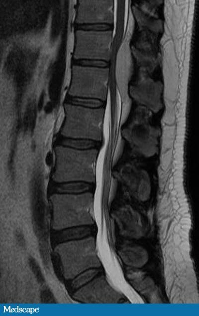 |
| Figure 1 |
On physical examination, his oral temperature is 98.6°F (37.0°C). His
pulse has a regular rhythm with a rate of 68 beats/min. His blood
pressure is 123/89 mm Hg. He is awake, alert, and oriented to time,
person, place, and situation. The cranial nerves II-XII are grossly
intact, and the patient's pupils are 3 mm and reactive to 2 mm. His
extraocular movements are intact. Face, tongue, uvula, and shoulder
shrug are symmetric. The motor examination of the lower extremities
reveals 5/5 strength in his bilateral hip flexors and knee extensors. He
has 4/5 strength with right dorsal and plantar flexion and 5/5 strength
with left dorsal and plantar flexion. Sensory examination reveals
intact sensation to vibration, but he has reduced pinprick sensation
from the midcalf and below bilaterally. Deep tendon reflexes are 1-2+,
with absent bilateral ankle clonus. He has an equivocal left Babinski
response with downgoing toes on the right. The patient's gait is normal.
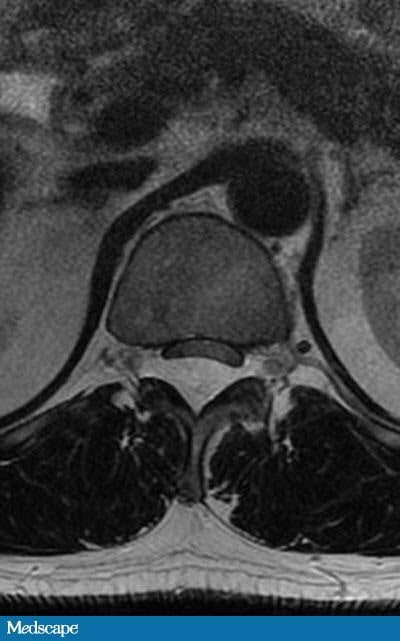 | |
| Figure 2 |
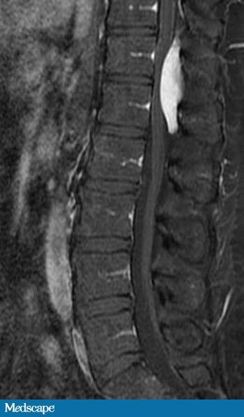 | |
| Figure 3 |
MR images with and without contrast of the thoracic and lumbar spine
are obtained, which reveal a posterior epidural mass involving the
T12-L1 level (Figures 1-3). Signal characteristics of the mass include
homogeneous isointense signal intensity on T1 (Figure 1), homogeneous
high signal on T2 (Figure 3), and uniform homogeneous postcontrast
enhancement (Figure 2). There is associated mass effect on the dorsal
aspect of the spinal cord without signal changes within the cord to
suggest edema or myelomalacia. The mass does not extend into the
adjacent neuroforamina nor does it involve the osseous structures and
the adjacent intervertebral disks, which have normal signal and
appearance. The paraspinal musculature and soft tissues have a normal
appearance. The visualized portions of the thorax and abdomen are
unremarkable. Laboratory analysis, including a complete blood count and a
basic metabolic panel, is normal.
What is the most likely diagnosis?
Hint: Note the location and the postcontrast characteristics of the mass.
Hint: Note the location and the postcontrast characteristics of the mass.
Epidural cavernous hemangioma
Epidural abscess
Ependymoma
Epidural hematoma
Tidak ada komentar:
Posting Komentar