BACKGROUND
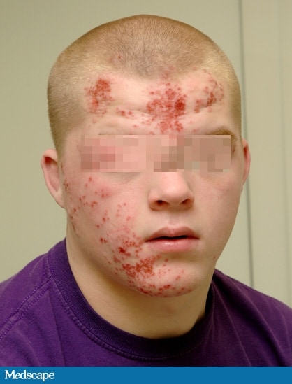 |
| Figure 1 |
A 17-year-old male high school student presents to the pediatric infectious disease clinic complaining of a 10-day history of a facial rash that "won't get better." The patient had previously visited his primary care provider (PCP), who started the patient on amoxicillin-clavulanic acid 8 days ago. The rash did not improve on the antibiotic, and as a result, it was discontinued and the patient switched to trimethoprim-sulfamethoxazole. No improvement was noted with the second round of antibiotic therapy; the rash continued to spread, and the lesions increased in number. The patient was subsequently advised to follow up with the infectious disease clinic.
At the infectious disease clinic, the patient states that the rash started with several pimples over the forehead and cheek and then continued to spread and involve most of the right side of his face. The lesions are not itchy, but they are painful. The patient has no known drug allergies. His immunizations are up to date. He is very active on the wrestling team and was happily preparing for an upcoming competition. The patient denies having any weight loss, headaches, dizziness, photophobia, fever, or chills. The family history is noncontributory.
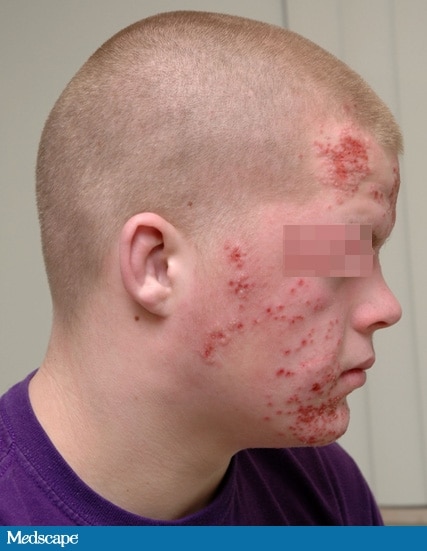 |
| Figure 2 |
On physical examination, the patient is alert and orientated. His oral temperature is 97.0°F (36.1°C). The patient has normal heart sounds, his pulse has a regular rhythm of 97 bpm, and his blood pressure is 125/75 mm Hg. His lungs are clear and his respiratory rate is 12 breaths/min. The examination of the head, eyes, ears, and nose is remarkable for multiple vesicular lesions measuring about 0.5 cm in diameter (see Figures 1 and 2). There is bilateral submandibular lymph gland enlargement measuring 1.5 × 1 cm. The neck is supple. His abdomen is soft and nontender to deep palpation in the epigastric region, and no organomegaly is noted. A complete blood count (CBC) taken at the PCP's office showed a white blood cell (WBC) count of 7.4 × 10³/µL (7.4 × 10⁹/L), with a normal differential; a hemoglobin of 13.6 g/dL (136 g/L); a hematocrit of 38.3% (0.3830); and a platelet count of 298 × 103/uL (298 × 109/L).
What is the most likely diagnosis?
BalasHapusHint: The patient was disqualified from wrestling because of his lesions.
#Tinea faciale
#Impetigo
#Bacterial cellulitis
#Herpes gladiatorum
i think it is herpes gladuatorum
BalasHapustinea faciale--> excluded with non-typical fungal lession which is central-healing lession
impetigo-->as far as i know it is most commonly in children and the lession is bullous and will make secondary crustose
bacterial cellulitis--> i think it will make some errotion or ulcus and leucosite rising mark ?
sorry i'm learning dermatovenerology in campuss now
cheers from jogja
Yes, you are correct. It is herpes gladiatorum.
BalasHapusIt is a viral infection which commonly occurs among wrestlers and other athletes who participate in close skin contact sports.
Regards from Semarang
The above described patient also complains of episodic eye pain, blurred vision, and sensitivity to light. What possible complication of herpes should you evaluate this patient for given these findings?
BalasHapus#Actinomycosis
#Pituitary apoplexy
#Ocular rosacea
#Dendritic keratitis